By
Skin Cancer Foundation •
Published On: April 27, 2026 •
Last Updated: April 28, 2026
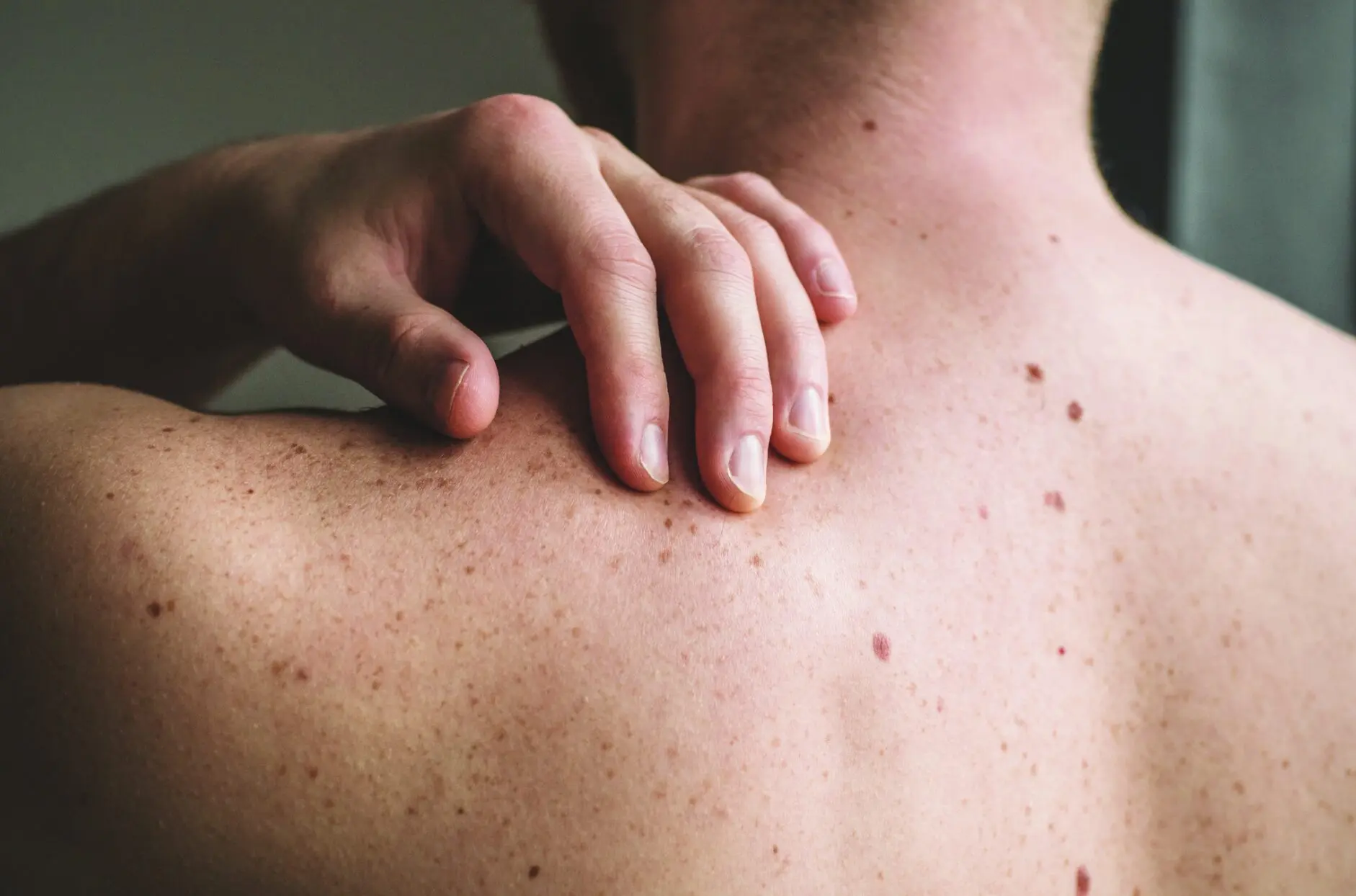
Early detection of melanoma can save your life. Look for anything new, changing or unusual on your skin and see your dermatologist regularly. Also check out the ABCDEs of melanoma for more information. Anastasiia Stiahailo / Getty Images
The goal of treating melanoma with Mohs surgery is the same as with nonmelanoma skin cancers: to examine 100 percent of the surgical margin and preserve as much healthy tissue as possible. With Mohs surgery, for both melanoma and nonmelanoma skin cancers, the surgeon and patient can be confident that the skin cancer is fully removed on the same day, before the wound is repaired.
Mohs surgery for melanoma can provide a high level of precision for early stage melanomas on cosmetically and functionally sensitive areas, such as the head and neck, hands, feet and genitals. To be more specific, Mohs surgery can be used to treat melanoma in situ (stage 0) and stage IA, while the tumor is confined to the upper layers of the skin and has no evidence of having spread. Mohs is not usually used for melanomas at stage IB or above. Those patients typically have a sentinel lymph node biopsy and excision of the melanoma performed in the operating room by a surgical oncologist.
The lab work required to examine the tissue under the microscope is distinctive in Mohs surgery, and the process of Mohs for melanoma is a little different than for nonmelanoma skin cancer. While the cells of BCCs and SCCs are easy to see under a microscope with standard frozen sections used in Mohs processing (see more on that, below), melanoma cells can be harder to detect. When Mohs surgery is used to treat melanoma, a staining process called immunohistochemistry (IHC) is performed to highlight the cancer cells on the slides. The stain highlights the melanocytes, which helps the surgeon see if cancer remains at the surgical margin. If so, the surgeon excises additional tissue in the precise area to remove the residual cancer.
Comparison of Mohs Surgery Versus Wide Local Excision
Traditionally, early melanoma has been treated with a wide local excision (WLE). For a WLE, the surgeon removes a football-shaped area around the original biopsy site, along with a margin of healthy-appearing skin. The size of the margin depends on the depth of the melanoma. The surgical site is then closed with stitches and bandaged. The patient goes home, and the tissue is sent to a lab where it is fixed in a chemical wax to form permanent sections for examination.
Once the tissue is fixed in wax, the lab technician vertically cuts the prepared tissue, like slicing a loaf of bread, perpendicular to the margins to look at representative sections of the removed region. It may take several days for results, and if more cancer cells are detected, the patient must return for additional surgery.
With Mohs surgery, the removed tissue is prepared differently. Frozen sections of the tissue are prepared in the lab on site by a technician and are sliced horizontally (en face), like layers of a cake, parallel to the margins. This allows the Mohs surgeon to view 100 percent of the margin under the microscope, which leads to higher cure rates and lower probability of recurrence.
Another Treatment Variation
There is also a variation of the WLE procedure for melanoma. You may have heard the term “slow Mohs” or “staged excision.” This is a method of excision where melanoma is excised and sent to the lab for permanent sections, and no wound closure is performed. This has the advantage of not repairing a wound without knowing that margins are clear, but the disadvantage of the process continuing over days to weeks. The surgeon removes the tissue, then the patient is carefully bandaged and sent home. The tissue is sent to a lab for processing and assessment. The patient returns to the surgeon for results and, if cancer cells remain, the surgeon repeats the procedure to remove the additional cancer. If the margins are clear, the surgeon closes the site.
For the high cure rate and low recurrence rate comparable to Mohs, staged excisions must be done with IHC stains and processed in a Mohs fashion, sliced horizontally (en face). I recommend a staged excision with en face margin assessment instead of a wide local excision with immediate closure for the treatment of early stage melanoma if you don’t have access to a provider who treats melanoma with Mohs surgery. This is especially important in cosmetically sensitive areas, like the face, where reconstructing these wounds may involve moving tissue from one area to an adjacent or distant area. Once such reconstruction is performed, there is no clear map of the positive margin in relation to the surgical site. This can make it more difficult for the surgeon to pinpoint the residual cancer if a second procedure needs to be performed.
The Bottom Line
Despite the benefits of Mohs surgery, it is not yet the standard of care for treating early stage melanoma. It requires special training for the Mohs surgeon and the histology technician. Cost and resources are another barrier to widespread adoption, as immunohistochemical stains are costly, with a short shelf life. It’s not a moneymaking procedure, but it is a service to patients that offers them the highest cure rate possible.
Nevertheless, the number of micrographic surgery and dermatologic oncology fellowship programs that include training on Mohs for melanoma are increasing, as are the number of Mohs surgeons performing the treatment. Promising research suggests the field will continue to grow. If you are a patient who has been diagnosed with early melanoma, you have options. Be your own advocate. Find a Mohs surgeon — and dermatologist — experienced in treating melanoma patients. And it’s never wrong to get a second opinion.
— Interview by Mia Medney
The patient, in his 70s, was referred to Dr. Lewin after his diagnosis of early stage melanoma on his right cheek. The biopsy area is visible as an ill-defined reddish-brown plaque, a good reminder that some melanomas can be very subtle and confused with “sun spots.” Dr. Lewin says pigmented lesions that have changed on sun-damaged skin should not undergo cosmetic procedures before checking for suspicion of skin cancer and/or performing a biopsy. This can save lives.
The patient needed only one stage of Mohs surgery with immunohistochemistry to remove the tumor before the results showed clear margins.
This type of reconstruction is called bilobed transposition flap elevated. It is favorable for skin cancer on the cheek area, allowing for careful placement of incisions to help camouflage the scar as it heals.
The closure, which slightly resembles a heart shape, will heal and flatten with time.
A later follow-up reveals the patient’s excellent healing and result.
Five medical photos courtesy of Dr. Jesse M. Lewin (written permission to share obtained from the patient).
About the Expert:
Jesse M. Lewin, MD, is an associate professor, director of The Kimberly and Eric J. Waldman Melanoma and Skin Cancer Center, vice chair of surgical operations, system chief of the Division of Dermatologic & Cosmetic Surgery and the program director for the Micrographic Surgery & Dermatologic Oncology Fellowship at The Kimberly and Eric J. Waldman Department of Dermatology at the Icahn School of Medicine at Mount Sinai in New York City.